Figure 1.
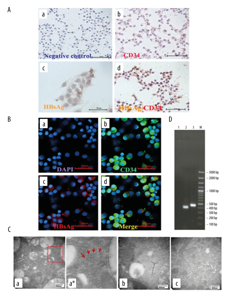
The infection of HBV in CD34-positive cells from cord blood. (A) Immunohistochemistry. (a) Negative control without primary antibodies; (b) CD34+ cells (red only) without HBV infection; (c) Expression of HBsAg (yellow) in HepG2.2.15 as positive control; (d) Expression of HBsAg (yellow) indicated in CD34+ (red) cells by DAB staining (×200). (B) Immunofluorescence. (a) Cell nuclei were visualized with DAPI; (b) CD34+ antigens (green) were found in membrane; (c) HBsAg (red) were located in the cytoplasm; (d) b and c were merged (×400). (C) Transmission electron microscopy. (a) HBV-infected CD34+ HSCs: HBV particles were roughly spherical with diameter of about 40 nm and localized exclusively in cytoplasm; (a*) Clipped and amplified HBV particles from Figure A; (b) HepG2.2.15 cells were used as positive control; (c) healthy CD34+ HSCs as negative control. (D) Reverse transcription-polymerase chain reaction (RT-PCR) products. Lane 1, healthy CD34+ HSCs as negative control; Lane 2, the 403 bp PCR products corresponding to the amplified HB S gene fragment; Lane 3, GAPDH as loading control.

