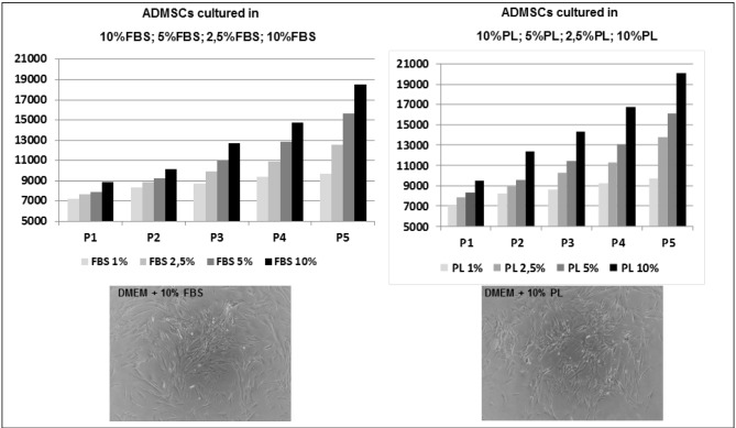
Figure 3.
Evaluation of the osteogenic potential of the PL.
Comparison of ADMSC cultured in standard conditions (left) and in the presence of PL (right). ADMSC had a comparable growth rate in the two media up to 12 passages. Different concentrations of FBS and PL were tested. Interestingly, there was no formation of any 3D multicellular spheroids as previously observed in SF/CBPG conditions. ADMSC: adipose mesenchymal stem cells; FBS: foetal bovine serum; PL: platelet lysate.

