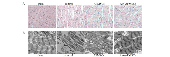
Figure 2.
Histological and ultrastructural effects of Akt-AFMSCs on myocardium in I/R rabbits. (A) Histological analysis was performed using hematox-ylin-eosin staining. Scale=100 µm. (B) Electron microscopic study of myocytes. Scale=500 nm. AFMSCs, amniotic fluid-derived mesenchymal stem cells; I/R, ischemia-reperfusion.

