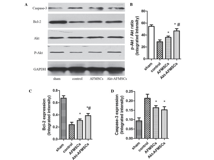
Figure 6.
(A) Protein expression of caspase-3, Bcl-2, Akt and P-Akt in hearts was assessed by western blotting. The Akt-AFMSC group showed an increased phosphorylation of (B) Akt (B) and (C) Bcl-2 in the left ventricle of rabbits subjected to myocardial ischemia-reperfusion. (D) The Akt-AFMSC and AFMSC groups showed a decreased level of caspase-3 in the left ventricle of rabbit subjected to myocardial ischemia-reperfusion. Data obtained from semiquantitative densitometry are presented as the mean ± standard deviation for eight rabbits per group. *P<0.05 vs. control; #P<0.05 vs. AFMSCs. Bcl-2, B-cell lymphoma 2; P, phosphorylated; AFMSCs, amniotic fluid-derived mesenchymal stem cells.

