Figure 7.
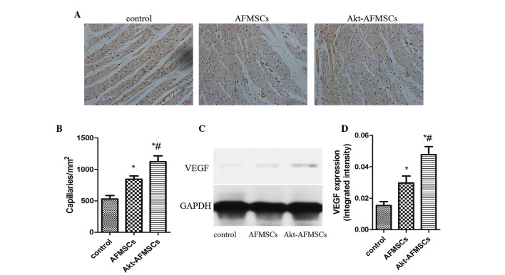
(A) Capillary density was measured using anti-von Willebrand factor immunohistology 21 days after transplantation. Blue staining shows the nuclei; brown staining shows the capillaries. Magnification, ×200. (B) The capillary density in the AFMSC and Akt-AFMSC groups was significantly higher than in the control group. *P<0.05 vs. control; #P<0.05 vs. AFMSCs. (C) VEGF secretion in recipient hearts was assessed by western blotting. (D) The levels of VEGF in the Akt-AFMSC group was significantly higher compared with that in control group. *P<0.05 vs. control; #P<0.05 vs. AFMSCs. GAPDH, glyceraldehyde-3-phosphate dehydrogenase; VEGF, vascular endothelial growth factor; AFMSCs, amniotic fluid-derived mesenchymal stem cells.

