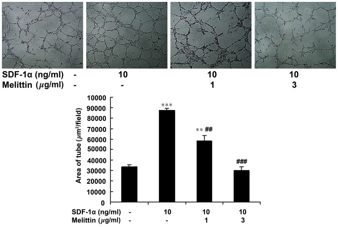
Figure 4.
Effect of SDF-1α and melittin on EPC tube formation ability. EPCs were divided into four groups: Control (untreated), 10 ng/ml SDF-1α, 1 µg/ml melittin + 10 ng/ml SDF-1α, and 3 µg/ml melittin + 10 ng/ml SDF-1α. Following 23 h of treatment, tube formation was determined (magnification, ×100). Data are presented as means ± standard deviation. **P<0.01, ***P<0.001 vs. controls; ##P<0.01, ###P<0.001 vs. SDF-1α. SDF-1α, stromal cell-derived factor-1α; EPC, endothelial progenitor cell.

