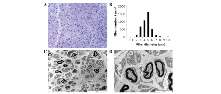
Figure 4.
Histopathological findings of the proband. (A) Semi-thin transverse sections revealed almost complete loss of large MFs with preserved medium- and small-sized MFs. Toluidine blue stain; magnification, x400. (B) Histogram demonstrates a unimodal distribution pattern of MFs with a diameter >8 μm, which comprised 0.9% of the total MFs. (C and D) Electron micrographs (magnification, ×3,000 and ×10,000, respectively) indicated that the remaining MFs had focal abnormalities of myelinated and unmyelinated axons, with occasional rarefaction. MFs, myelinated fibers.

