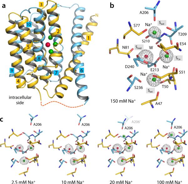
Figure 1.
Na+ binding to outward-facing NCX_Mj. (a) Overall structure of native outward-facing NCX_Mj from crystals grown in 150 mM Na+. N- and C-terminal halves are colored yellow and cyan, respectively. Colored spheres represent the bound Na+ (green) and water (red). (b) Structural details and definition of the four central binding sites. Only residues flanking these sites are shown for clarity (same for all other figures). The electron density (grey mesh, 1.9 Å Fo-Fc ion omit map contoured at 4σ) at Smid was modeled as water (red sphere) and those at Sext, SCa and Sint as Na+ ions (green spheres). Further details are shown in Supplementary Fig. 1. (c) Concentration-dependent change in Na+ occupancy (see also Table 1). All Fo – Fc ion-omit maps are calculated to 2.4 Å and contoured at 3σ for comparison. The displacement of A206 reflects the [Na+]-dependent conformational change from the partially open to the occluded state (observed at low and high Na+ concentrations, respectively). At 20 mM Na+, both conformations co-exist. No significant changes were observed in the side-chains involved in ion or water coordination at the SCa, Sint and Smid sites.

