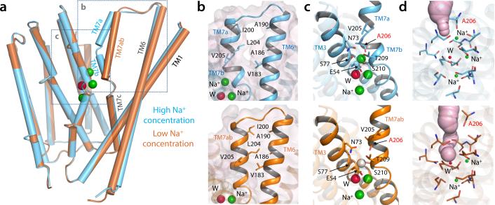
Figure 2.
Na+-occupancy dependent conformational change in NCX_Mj. (a) Superimposition of the NCX_Mj crystal structures obtained in high (100 mM, cyan cylinders) and low (10 mM, brown cylinders) Na+ concentrations. (b) Close-up view of the interface between TM6 and TM7ab in the NCX_Mj structures obtained at high and low Na+ concentrations (top and lower panels, respectively). Residues forming van-der-Waals contacts in the structure at low Na+ concentration are shown in detail. (c) Close-up view of the Na+-binding sites. The vacant Sext site in the structure at low Na+ concentration is indicated with a white sphere. Residues surrounding this site are also indicated; note A206 (labeled in red) coordinates Na+ at Sext via its backbone carbonyl oxygen. (d) Extracellular solvent accessibility of the ion binding sites in the structures at high and low [Na+]. Putative solvent channels are represented as light-purple surfaces.

