Figure 3.
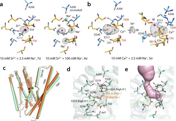
Divalent cation binding and apo structure of NCX_Mj. (a) A single Sr2+ (dark blue sphere) binds at SCa in crystals titrated with 10 mM Sr2+ and 2.5 mM Na+ (see also Supplementary Fig. 2). Residues involved in Sr2+ coordination are labeled. There are no significant changes in the side-chains involved in ion coordination, relative to the Na+-bound state. T50 and T209 (labeled in red) coordinate Sr2+ through their backbone carbonyl-oxygen atoms. High Na+ concentration (100 mM) completely displaces Sr2+ and reverts NCX_Mj to the occluded state (right panel). The contour level of the Fo – Fc omit map of the structure at high Na+ concentration was lowered (to 4σ) so as to visualize the density from Na+ ions and H2O. (b) Ca2+ (tanned spheres) binds either to SCa or Smid in crystals titrated with 10 mM Ca2+ and 2.5 mM Na+ (see also Supplementary Fig. 2). The relative occupancies are 55% and 45%, respectively. (c) Superimposition of NCX_Mj structures obtained at low Na+ concentration (10 mM) and pH 6.5 (brown) and in the absence of Na+ and pH 4 (light green), referred to as apo state. (d) Close-up view of the ion-binding sites in the apo (or high H+) state. The side chains of E54 and E213 from the low Na+ structure are also shown (light brown) for comparison. White spheres indicate the location Sint, Smid SCa. (e) Extracellular solvent accessibility of the ion-binding sites in apo NCX_Mj.

