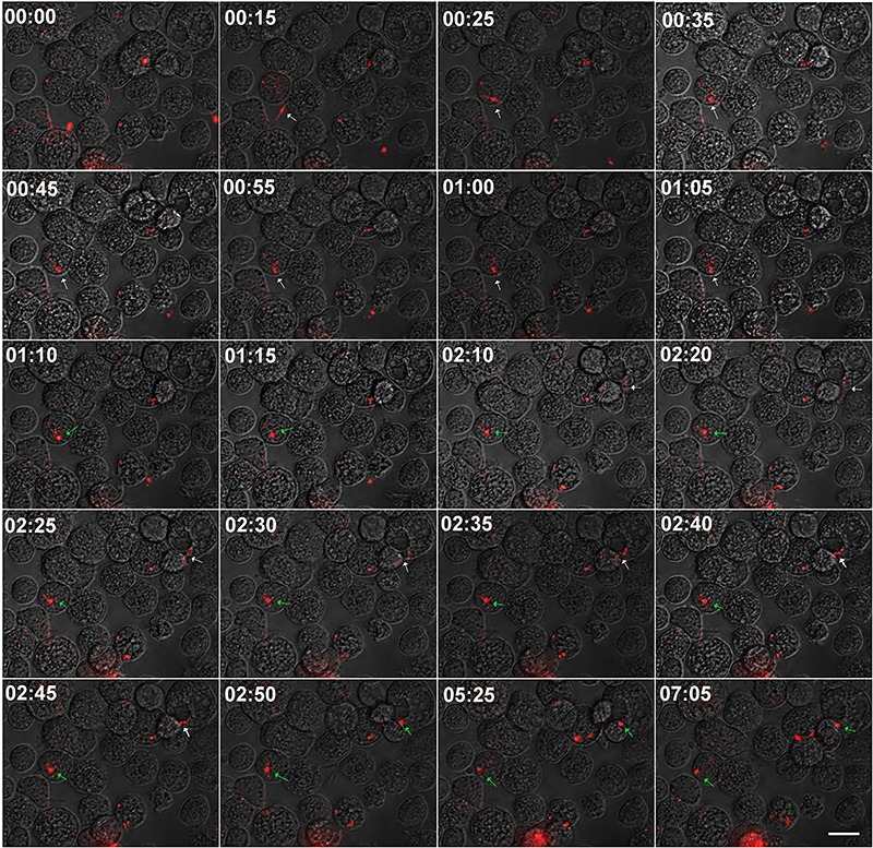
Figure 4. Time-lapse analysis of Borrelia burgdorferi uptake by AVL/CTVM17 cells. PKH26 stained B. burgdorferi were added at a multiplicity of 50 bacteria to each tick cell to a culture of AVL/CTVM17 cells and monitored by time-lapse microscopy during the first 7 h, with a 5 min interval. The selected images show an extracellular B. burgdorferi (white arrow) taking about 50 min to attach to and be internalized by a tick cell (green arrow). The scale bar represents 25 μm.

