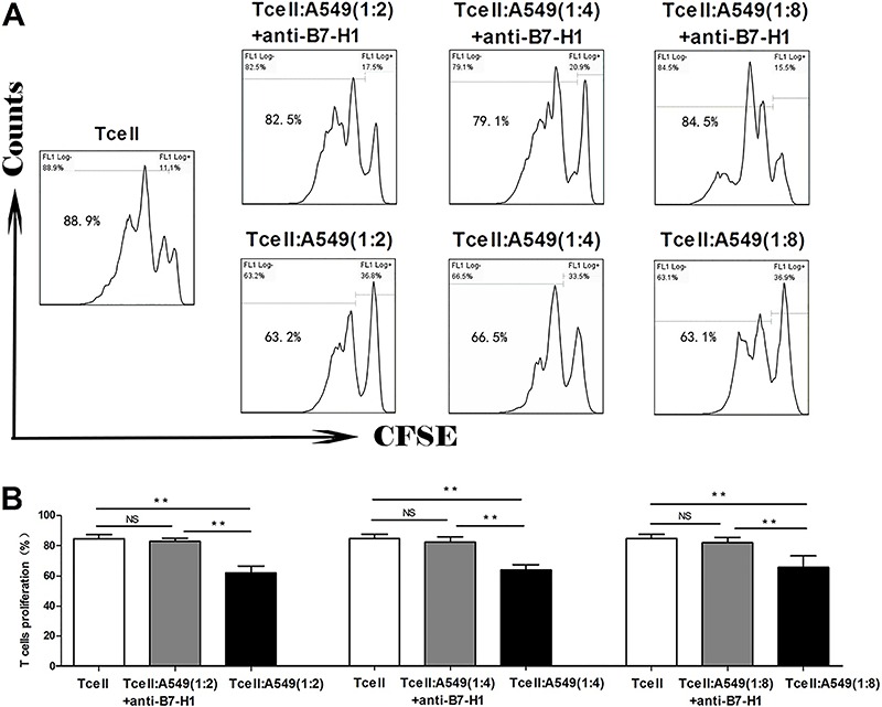
Figure 1. A, Representative flow cytometry graphs for each experimental group, with the percentage of proliferating T cells (cells left to the first peak of carboxyfluorescein succinimidyl ester (CFSE)+ cells) labeled in the graph. B, Quantitative analysis of proliferating T cells among different groups. Tumor-associated B7-H1 is essential for regulating T-cell proliferation. T cells isolated from human peripheral blood mononuclear cells were labeled with CFSE, stimulated with anti-CD3 and anti-CD28, and co-cultured with T cells alone or with A549 cells at the T:A549 ratios of 1:2, 1:4 or 1:8, in the absence (+A549) or presence of anti-B7-H1 (+anti-B7-H1). T-cell proliferation was examined by flow cytometry after 72 h. NS: non-significant. **P<0.01 (ANOVA).