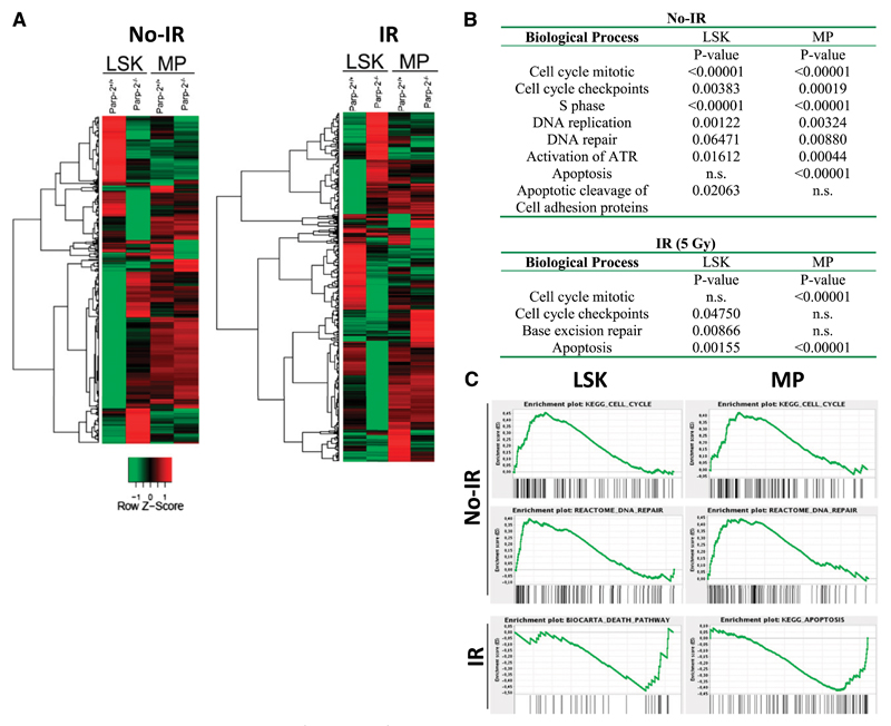
Figure 7. Gene expression profile analysis of Parp-2+/+ and Parp-2−/− LSK and MP cells.
(A) Heat map representing the average normalized intensity values of all genes that are differentially expressed between WT and Parp-2−/− LSK and MP cells (n = 982 genes). Red indicates higher expression in Parp-2−/− compared with Parp-2+/+, whereas green indicates lower expression in Parp-2−/− compared with Parp-2+/+. (B) Selected differentially expressed biological processes between WT and Parp-2−/− LSK and MP cells. Canonical pathways gene sets were scored using the GSEA and P values were computed using 1000 permutations. n.s., not statistically significant. (C) Examples of GSEA plots obtained from expression microarray data. Within these plots, the green line represents the sliding enrichment score and the black bars demarcate the position of the gene set members within the ranked expression data.

