Fig. 3.
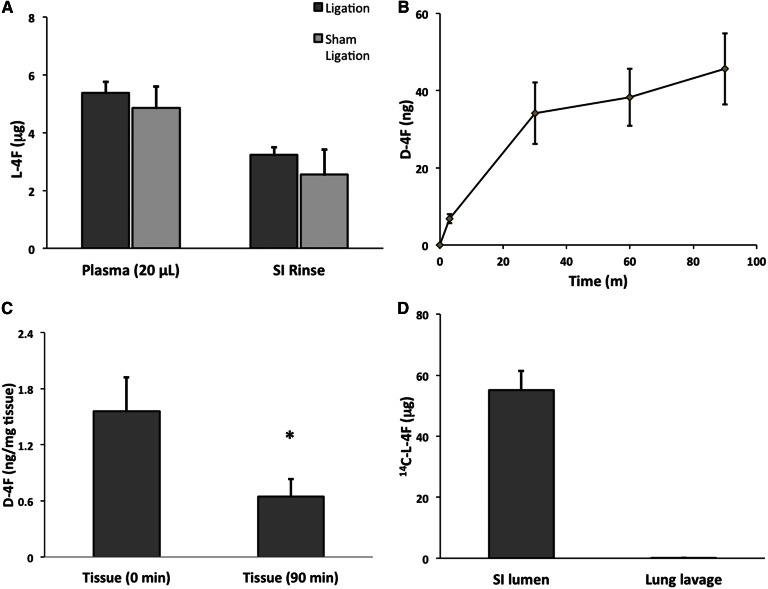
l-4F enters the intestinal lumen by directly and selectively crossing the intestinal epithelium. A: l-4F (25 mg/kg) was introduced via tail vein into C57BL/6J mice, whose bile ducts had either been ligated or sham ligated (n = 4). Intact l-4F present in the plasma and SI PBS rinse after 1 h was quantified by LC/MS/MS. B, C: C57BL/6J mice (n = 7) were injected with 800 μg (∼25–30 mg/kg) of d-4F via tail vein. After 30 min, the mice were euthanized, and duodenal explants were mounted in Ussing chambers. Serosal side media contained LDL and HDL, while mucosal side media included micelles. Mucosal side media was sampled at 3, 30, 60, and 90 min; and the amount of d-4F present was determined by LC/MS/MS. Peptide was observed released from the tissue into the mucosal side media across 90 min (B). Levels of d-4F were also quantified in these duodenal explants together with matched controls from the same mice. The controls were not mounted in the Ussing chamber and provide the baseline or time zero concentration of d-4F in the tissue (C). Ussing chamber mounted tissue after 90 min had significantly less d-4F than controls (* P < 0.05). D: In order to determine whether l-4F selectively crosses intestinal epithelium, the level of l-4F present in lung alveolar space was determined via lung lavage following two individual 25 mg/kg injections of 14C-l-4F at 1 and 3 h prior to being euthanized (n = 6). No l-4F was present in the lung lavage, in contrast with SI PBS rinse. The dpm were converted to μg l-4F. Error is reported as SEM.

