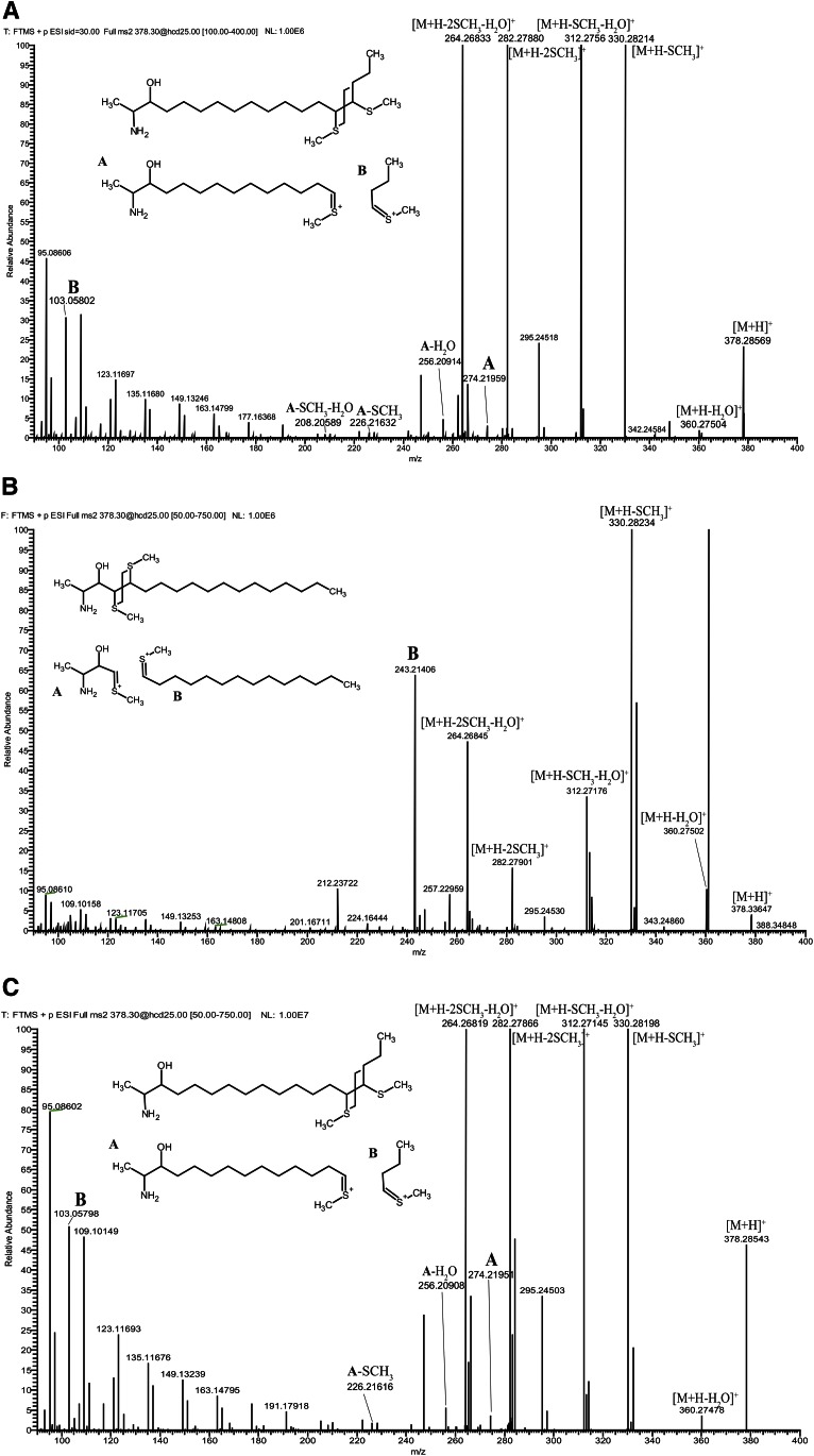
Fig. 2.
A: Hydrolyzed lipid extract from HEK293 cells was subjected to DMDS derivatization and analyzed on a high-resolution accurate mass spectrometer using ESI and direct injection. The [M+H]+ ion formed from DMDS adduct of native 1-deoxySO (m/z of 378.2857) was in excellent agreement (<1 ppm) with the calculated m/z of 378.2859 (C20H44NOS2+). The precursor ion was selected for CID yielding to the spectrum shown. Product ions A (m/z 274) and B (m/z 103) are indicated in the spectrum and are diagnostic for a fragmentation at (Δ14) position. B: Spectrum from SPH m18:1(4E)(3OH) showing product ion B (m/z 243). C: Spectrum from SPH m18:1(14Z)(3OH) showing product ions A (m/z 274) and B (m/z 103). Putative structures for the diagnostic product ions are shown as insets in the spectra.

