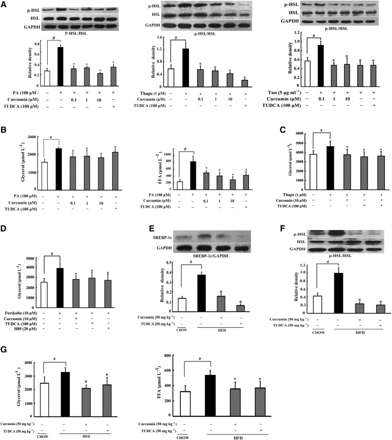
Fig. 3.
Curcumin inhibited adipose lipolysis. A: HSL phosphorylation in adipose tissue subjected to PA or thapsigargin (Thaps) challenge (24 h) were determined by Western blot. B: Glycerol and FFA release in adipose tissue treated with PA. C, D: Glycerol release in adipose tissue exposed to Thaps and forskolin for 24 h (n = 6). SREBP-1c (E) and p-HSL/HSL (F) in adipose tissue of HFD-fed mice were determined by Western blot. G: Glycerol and FFA contents in the AT-CM of HFD-fed mice (n = 6). Data are expressed as the mean ± SD of four independent experiments. *P < 0.05 versus model; #P < 0.05 versus control.

