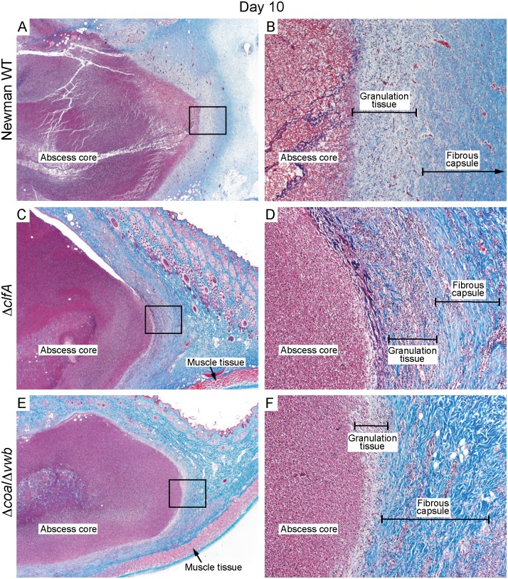
Fig 2. Histopathology of rabbit skin abscess caused by S. aureus.
Histopathology sections represent skin abscesses caused by S. aureus Newman WT (A, B), ΔclfA (C, D) or Δcoa/Δvwb (E, F) strains on day 10 post-infection. Abscess sections were stained with standard Masson’s trichrome stain to enhance fine structure detail of muscle tissues, collagen fibers and fibrin. (A, C and E) original magnification is 20×. (B, D, and F) 200× magnification of selected area (black rectangle) depicted in panels A, C or E.

