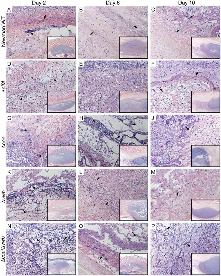
Fig 3. Fibrin deposition in rabbit skin abscess caused by S. aureus Newman.
Representative sections of rabbit skin abscesses on Day 2 (A, D, G, K, N), Day 6 (B, E, H, L, O) and Day 10 (C, F, J, M, P) post infection. Abscesses from rabbits infected with S. aureus Newman WT (A-C), ΔclfA (D-F), Δcoa (G-J), Δvwb (K-M) and Δcoa/Δvwb (N-P). Tissue sections were stained with Mallory’s phosphotungstic acid-hematoxylin stain for visualization of fibrin (black arrows). Magnification is 200×. Inset image is the abscess at 20× (black rectangle).

