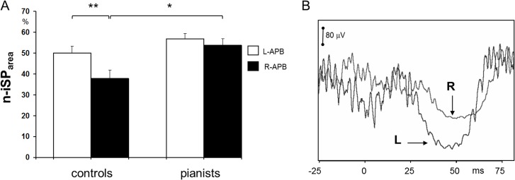
Fig 5.
A. Average normalized ipsilateral silent period area (n-iSParea) on the left and right APB in pianists and naïve controls, the latter showing a wake suppression of voluntary EMG on the right APB to stimulation of the non-dominant ipsilateral hemisphere (error bars: standard error of the mean. Left vs right APB in controls **p<0.001; controls vs pianists R-APB *p = 0.008). B. Example of ipsilateral silent period on right APB (R) and left APB (L) in a control naïve subject. Note the stronger inhibition of voluntary EMG on the left, non-dominant side by stimulating the ipsilateral left, dominant hemisphere.

