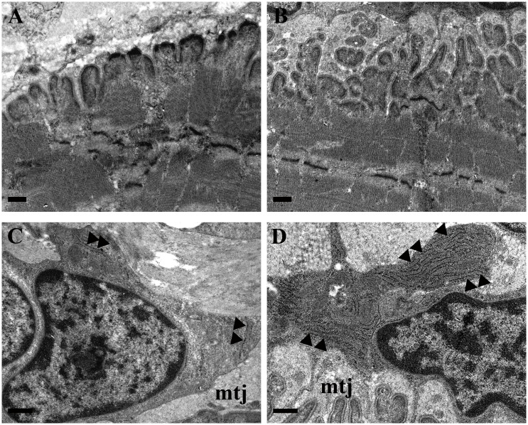
Fig 3. Morphological features of MTJ in control and trained rats.
TEM images of MTJs which reveal the different complexity between control (A) and trained groups (RUN-F, B). At high magnification (13500x), tenocytes, located strictly close to MTJ (mtj), display, in comparison to control (C), a growing presence of rough endoplasmic reticulum (►) in trained groups (RUN-F, D). Bars A,B,C,D: 0.5μm.

