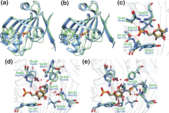
Fig 9. Effector recognition is highly similar in DasR and NagR.
(a) and (b) Superposition of DasR-EBD (blue) and NagR (green; truncated to NagR-EBD) in complex with the phosphorylated sugars GlcN-6-P (a) and GlcNAc-6-P (b). The r.m.s. deviations of the Cα-positions of the superimposed structures are (a) 1.39 Å (a) and (b) 1.47 Å. For clarity, only the protein chain that is coordinating the α-anomer of GlcN-6-P or GlcNAc-6-P in the dimeric repressor is displayed. The phosphorylated sugars are depicted as stick models and coloured in grey (bound by DasR) or gold (bound by NagR). (c) Interaction of the phosphate moiety of GlcN-6-P with DasR and NagR. (d) and (e) Interaction of the sugar moiety of GlcN-6-P (d) and GlcNAc-6-P (e) with DasR and NagR. Protein residues are presented as stick models. Water molecules are shown as red spheres.

