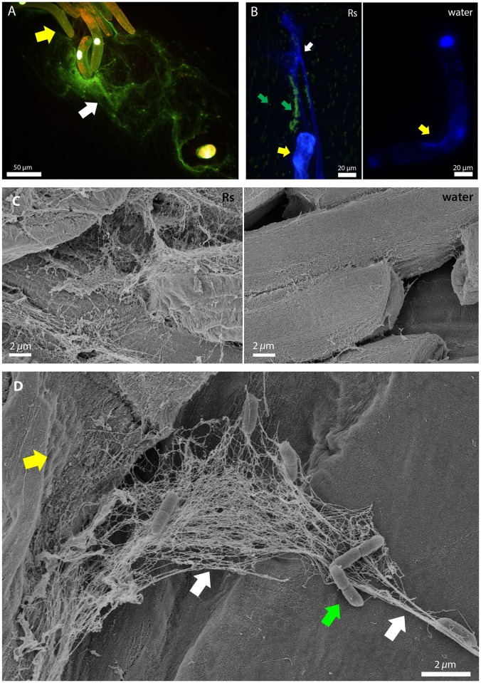
Fig 1. Plant root border cells release DNA-containing extracellular traps in response to R. solanacearum.
(A) Fluorescence microscopy images of pea root border cells (yellow arrow) releasing extracellular DNA (white arrow, stained green or white with SYTOX Green) 30 min after exposure to R. solanacearum cells. (B) A pea root border cell (yellow arrow) treated with GFP-expressing R. solanacearum (green arrows), which are immobilized on traps containing extracellular DNA (stained blue with DAPI, white arrow). In the adjacent water control, the root border cell nuclei are stained blue but no extracellular DNA is visible. Untrapped bacteria were able to move freely in the suspension and appear blurred in the image. (C) Scanning electron microscopy showed that following treatment with R. solanacearum, pea border cells released web-like structures similar to neutrophil extracellular traps. (D) Root extracellular traps contained both small threads and thicker cables (left and right white arrows, respectively). R. solanacearum cells (green arrow) were captured by traps originating from a collapsed pea root border cell (yellow arrow).

