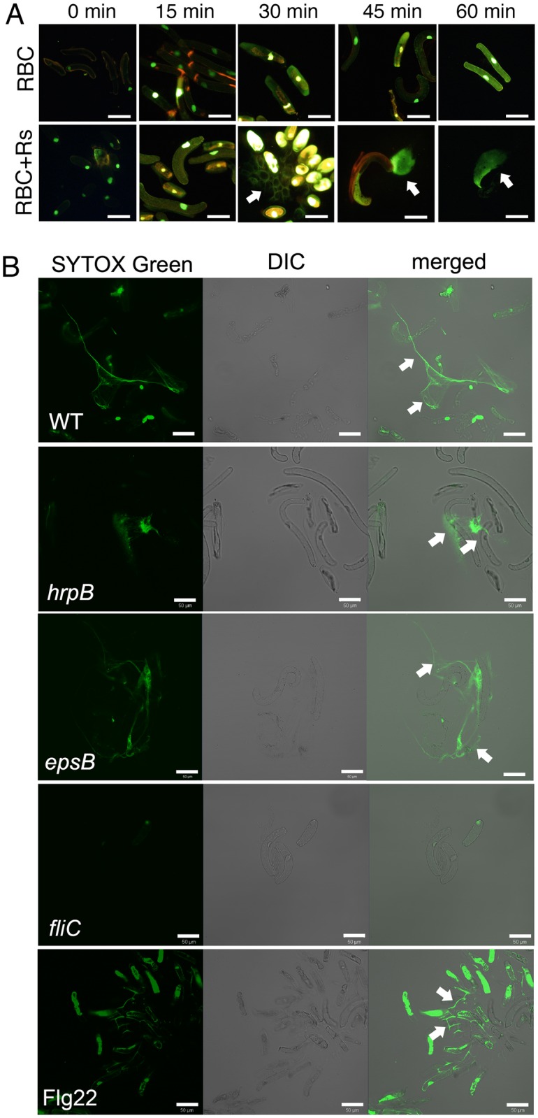
Fig 2. Flagella and Flg22 triggered the release of exDNA by pea border cells.

(A) Kinetics of root border cell extracellular trap release. Pea root border cells (RBC) were inoculated with 106 cells of R. solanacearum strain GMI1000 (Rs), the preparation was stained with SYTOX Green, and imaged over time with an epifluorescence microscope. Images are representative of two experiments and at least four images were taken for each time point. (B) Flagellin-triggered formation of border cell extracellular traps. A suspension of 10,000 pea border cells were inoculated with a 1000-fold excess (107 cells) of either R. solanacearum wild-type strain GMI1000 (WT), type III secretion system mutant hrpB, exopolysaccharide deficient mutant epsB, flagellin deficient mutant fliC, or 20 μg/ml of flagellin-derived peptide Flg22 and stained with SYTOX Green to visualize DNA (white arrows on merged images). Live imaging was performed with a Zeiss Elyra 780 CLSM and SYTOX Green fluorescent, differential interference contrast microscopy (DIC) and merged images are shown. At least 5 images per treatment were taken from 30 min to 1 h post inoculation. The experiment was repeated three times and representative images are shown (bar = 50 μm). White arrows in (A) and (B) indicate exDNA.
