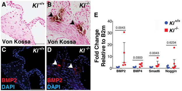
Figure 3. BMP pathway components are active in calcified Klotho−/− AoV.
BMP2 ligand is expressed in calcified AoV of Klotho−/− mice (B,D) but not control AoV (A,C) at 6 weeks of age, as determined by immunofluorescence (red). BMP signaling network gene expression was evaluated by qRT-PCR of RNA isolated from Klotho−/− AoV compared to WT controls at 6 weeks of age (E). There is a significant increase in BMP2 and BMP4 ligand mRNA levels, as well as Smad6 (n=5/group). Gene expression was normalized to B2m control expression. Values were normalized to wild type control averages and are represented as fold changes. Error bars are shown as interquartile means and scatter plot. Statistical significance was determined by Mann-Whitney U-test (p< 0.05).

