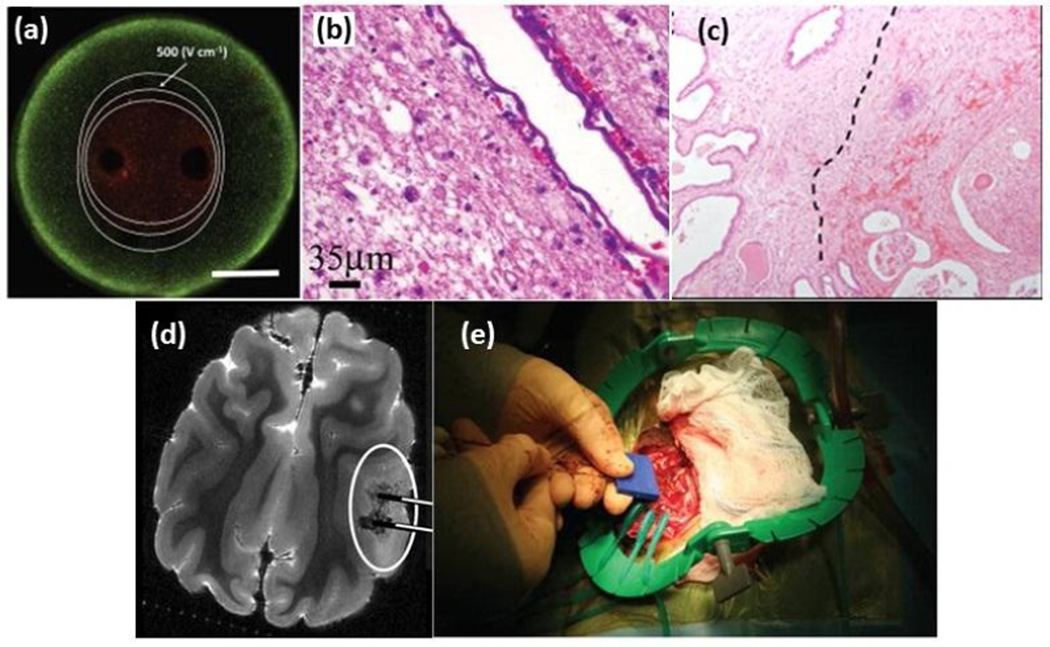
Figure 2.
Irreversible electroporation from in vitro to in vivo (a) visualization of live/dead cells after IRE treatment in a 3D in vitro tumor model [101], (b) Sparing of major blood vessels after IRE treatment of canine brain [102], (c) Delineation between viable tissue (left) and reactive fibrosis and hemorrhage (right) is seen in human prostate IRE histology, adapted from [103], (d) 7.0-T MRI of IRE-treated canine brain, adapted from [104], (e) dual probe insertion during the intracranial IRE procedure in canine brain [102]
Owing to their short rise time which is faster than cell membrane charging time, nanosecond pulsed electric fields (nsPEFs) penetrate the cell membrane and damage intracellular structures, with minimal membrane electroporation [105, 106]. In vitro studies on skin cells have shown that tumor cells have a stronger response to nsPEFs than normal cells [107, 108], and suggest that an improved understanding of the electrical differences among cells may lead to more targeted therapies based on exploiting these differences.

