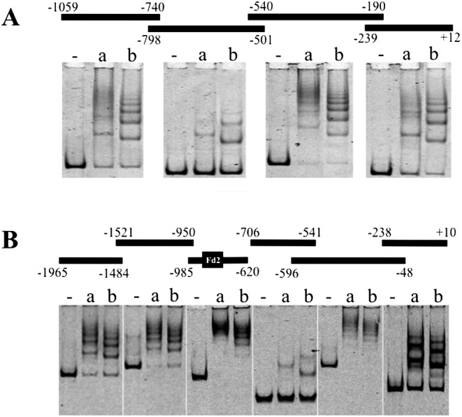
Fig. 2.

Electrophoretic gel mobility shift assay of LHX2 and LHX3. Upstream fragments of Cga (A) and Fshβ (B) are indicated with a line, (thick bar in B indicates Fd2) and nucleotide number above the electrogram. The mixture without protein (-) or with recombinant LIM domain-deleted LHX2 (ΔLIM-LHX2; a) and LHX3 (ΔLIM-LHX3; b) proteins and FAM-labeled fragments were analyzed on a 4% polyacrylamide gel followed by visualization with a fluorescence viewer.
