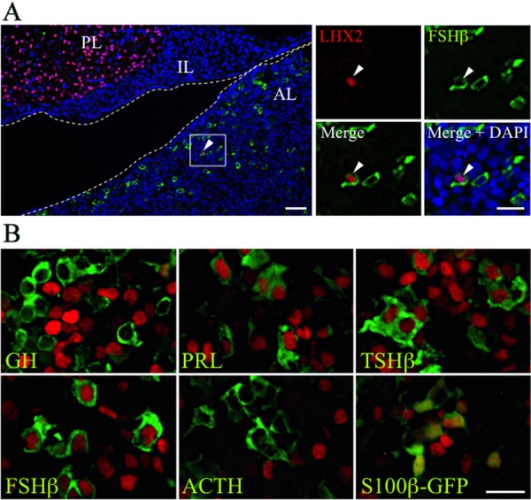
Fig. 5.

Immunohistochemistry of LHX2 and LHX3 in the postnatal pituitary. (A) Double-immunostaining of LHX2 (Cy3, red) and FSHβ (Cy5, green) for section on P15 was performed. Merged image with DAPI (nuclei) is shown in the left panel and boxed areas are enlarged in the right panels. Arrowheads indicate LHX2/FSHβ-double positive cells. Dotted lines indicate Rathke’s cleft. AL, anterior lobe; IL, posterior lobe; PL, posterior lobe. Bars in the left and right panels indicate 50 and 20 µm, respectively. (B) Double-immunostaining of LHX3 (Cy3, red) and pituitary hormones (Cy5, green with false color) or S100β-GFP (FITC, green) for section on P15 were performed. Bar indicates 20 µm.
