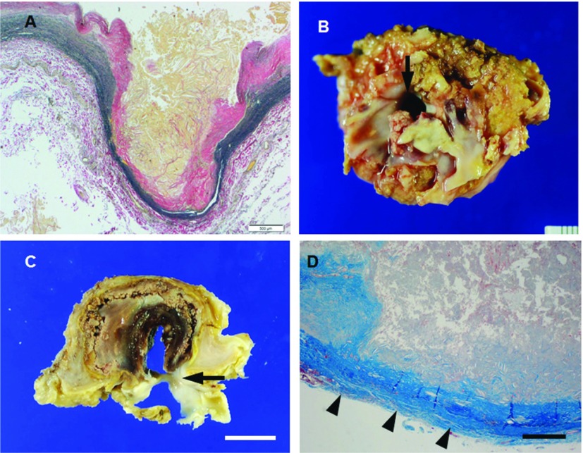
Fig. 1.
(A, B) Microphotograph of the abdominal aortic wall section stained with elastic van Gieson from patient (69 y.o., male) with atherosclerotic aneurysm. Atheromatous lesion extends into the media with loss of elastic fibers, the abdominal aortic wall is thinned and results in aneurysm. (B–D) Macrophotographs (B, C) and microphotograph (D, Masson’s trichrome) of the thotacic aorta from patient (79 y.o., female) with PAU. Ulceration of atherosclerotic plaque that contact to the adventitia (arrowheads in D) with intimal disruption (arrows in B and C) of the thoracic aorta, leading to subadventitial pseudoaneurysm. Scale bars: Panel A: 500 µm; Panel C: 1 cm; Panel D: 200 µm. PAU: penetrating atherosclerotic ulcer

