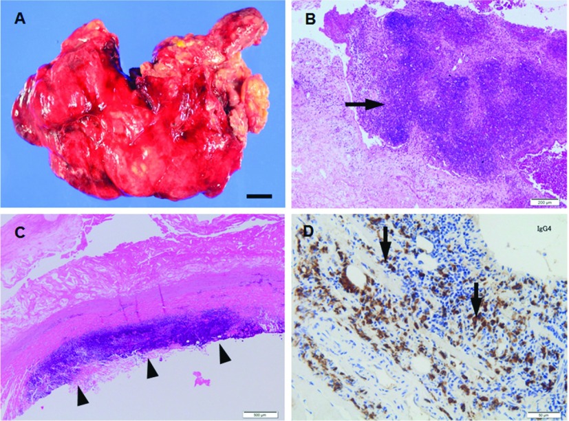
Fig. 2.
(A, B) Macrophotograph (A) and microphotograph (B, H.&E.) of the abdominal aortic wall from patient (69 y.o., male) with infected aortic aneurysm. Aortic aneurysm shows saccular form aneurysm with irregular configulation (A) and neutrophils infiltrate with medial destruction (arrow in B). (C, D) Microphotographs (C, H.&E.; D, immunohistochemistry for IgG4) of the abdominal aortic wall sections from patient (69 y.o., male) with IAAA. Fibrosing adventitia shows lymphoplasmacytic infiltrate that appears as a bands and clusters (C). Large population of plasma cells shows positive reactivity for IgG4 (D). Scale bars: Panel A: 1 cm; Panel B: 200 µm; Panel C: 500 µm; Panel D: 50 µm. IAAA: Inflammatory abdominal aortic aneurysm

