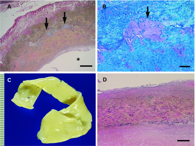
Fig. 4.
(A, B) Microphotographs (A, elastica van Gieson; B, toluidine blue) of the thoracic aortic wall sections from patient (30 y.o., male) with MFS. The media shows cystic medial degeneration with proteoglycan deposits and elastic fiber fragmentation of the aorta (arrows in A, B). (C, D) Macrophotograph (C) and microphotograph (D, elastica van Gieson) of thoracic aortic wall sections from patient (23 y.o., male) with LDS. Aortic wall is nonatherosclerotic and its media shows diffuse medial degeneration with elastic fiber fragmentation. Scale bars: Panel A: 500 µm; Panel B: 100 µm; Panel D: 500 µm. * represents vascular lumen. MFS: Marfan syndrome; LDS: Loeys-Dietz syndrome

