Fig. 5.
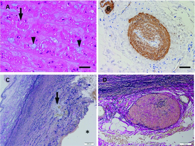
(A, B) Microphotographs (A, H.&E.; B, immunohistochemistry for a-smooth muscle actin) of the thoracic aortic wall sections from patient (50 y.o., male) with ACTA2 mutation. The aortic wall shows disarrangement of vacuolated (arrow in A) of SMCs with abundant extracellular matrix (arrowheads in A) in the media and vasa vasorum with smooth muscle cell hyperplasia (FMD) in adventitia (B). (C, D) Microphotographs (C, elastica van Gieson; D, elastica van Gieson) of the ascending aortic wall sections from patients (51 y.o., male; 56 y.o., male) with MYH11 mutation. The aortic wall shows elastic fiber fragmentation with cystic medial degeneration in the media (arrow in C) and vasa vasorum with medial thickening (FMD) in the adventtiia (D). Scale bars: Panel A: 25 µm; Panel B: 50 µm; Panel C: 200 µm; Panel D: 100 µm. * represents vascular lumen. SMC: smooth muscle cell; FMD: fibromuscular dysplasia

