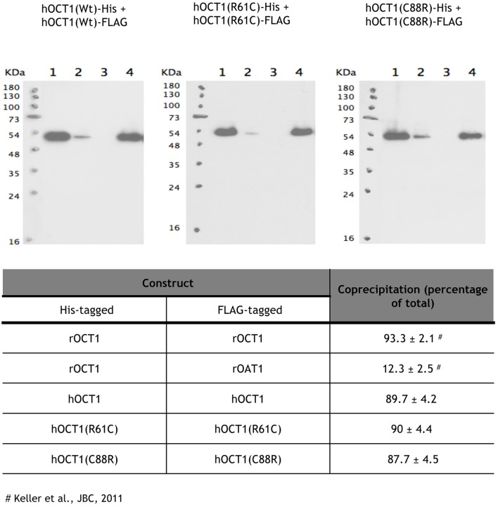
Figure 2.
Oligomerization of polymorphic variants located at the extracellular loop. The samples were diluted with 200 μL of Tris buffer containing 1% CHAPs and 10 mM imidazole (lanes 1). After 1 h incubation at 4°C, Ni2+-NTA-agarose beads were added, the suspension was incubated for 1 h and centrifuged. Supernatants were collected (lanes 2). The beads were washed five times with 1 mL of buffer and pelleted (supernatants lanes 3). His-tagged proteins were eluted by incubating the beads with 400 μl of buffer containing 1% CHAPS and 100 mM imidazole (supernatant lanes 4). Proteins were separated by SDS-PAGE, transferred to a blotting membrane, and stained with an antibody against the FLAG tag. The experiment was performed three independent times. The mean ± S.D. are shown in the table.

