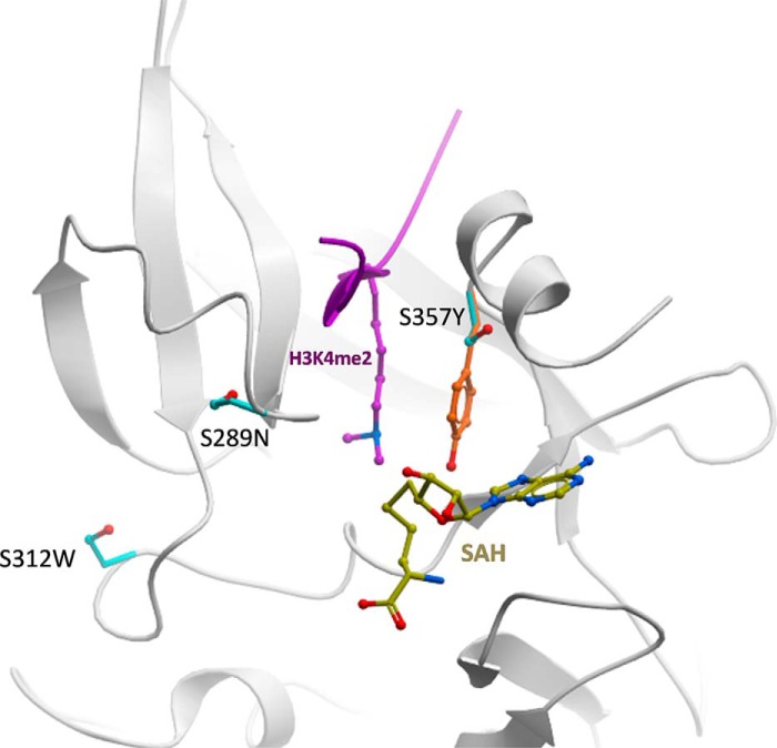
FIGURE 7.
Mapping of non-conserved side chains on a structural model of PRDM7. The three PRDM7 serine side chains (cyan) that are not conserved in PRMD9 are mapped on a model of PRDM7 (white ribbon) built by homology to a mPRDM9 structure in complex with S-adenosyl-l-homocysteine (SAH) (yellow) and a H3K4me2 peptide (magenta). The catalytic tyrosine of PRDM9 (orange) is shown.

