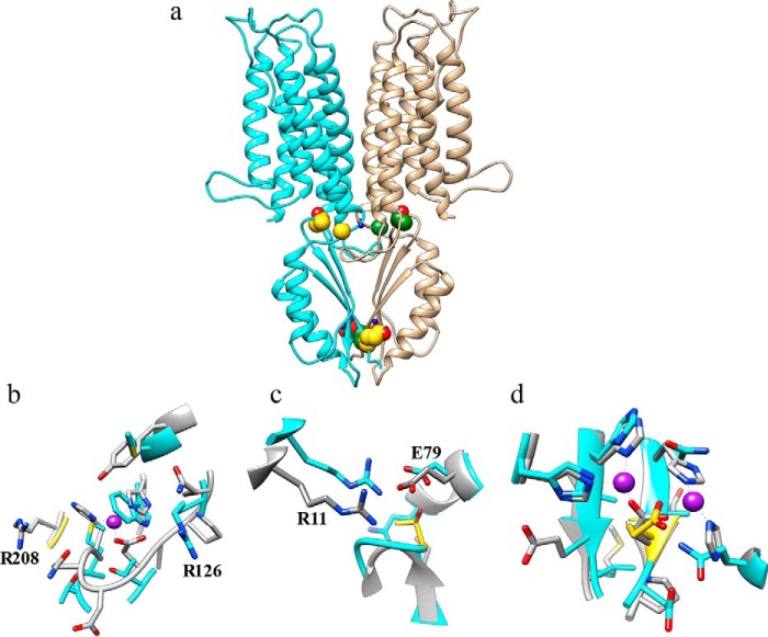
FIGURE 6.
Three-dimensional structural modeling of the human ZnT2. The ZnT2 monomer model was aligned to the PDB code 3h90 template by the HHpred method as described under ”Materials and Methods.“ (a) monomer ribbons are colored in cyan and tan, respectively, and the positions of the various ZnT2 mutations are shown in CPK (Corey-Pauling-Koltun) representation (available in Chimera), where carbon atoms are colored in gold and green for the two different monomers, respectively. Zinc ions from the YiiP model are shown (for approximate location only) in blue CPK balls. (b) the Microenvironment of Gly-280 in the human ZnT2 model; the carbon atoms of Gly-280 are colored in gold. (c) The environment of Thr-312 in the human ZnT2 model. (d) The environment of Glu-355 in the human ZnT2 model; the proximity to the two zinc atoms from the YiiP crystal structure and the two histidine residues (which are also present on YiiP), strongly suggests that Glu-355 is directly involved in zinc coordination. The mutation sites are colored in gold. Carbons of the ZnT2 model colored in cyan and carbons of the YiiP colored in gray. Oxygen atoms are colored in red and nitrogen atom in blue. Zinc atoms are colored in violet.

