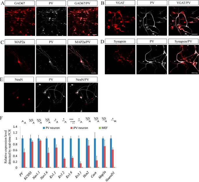
FIGURE 2.
iPV neurons express the specific markers of PV neurons. A and B, at 14 days post-infections, MEF-derived iN cells expressed two specific inhibitory neuronal markers of GAD67 (A) and VGAT (B). C–E, at 10–14 days post-infection, iPV neurons expressed the pan-neuronal markers Map2a (C), synapsin (D), and NeuN (E) along with PV. F, real-time fluorescence quantification PCR showed that neuronal or PV-specific genes were up-regulated in iPV neurons, and this tendency is consistent with that in endogenous PV neurons. *p < 0.05, **p < 0.01, ***p < 0.001. NS (not significant), p > 0.05. Scale bars: 50 μm.

