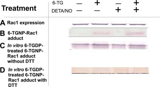
FIGURE 3.

DETA/NO does not enhance formation of the 6-TGNP-Rac1 disulfide adduct in CD4+ cells. A, Western analysis of CD4+ cells for the determination of Rac1 expression was performed using an anti-Rac1 antibody as described under “Experimental Procedures.” B, presence of the 6-TGNP-Rac1 disulfide adduct in the IPed Rac1 from CD4+ cells was examined by Western analysis by using an anti-6-TGNP antibody as noted under “Experimental Procedures.” C, Vav-mediated formation of the 6-TGNP-Rac1 disulfide adduct in the IPed Rac1 from CD4+ cells in the presence of 6-TGDP and/or NO was analyzed by Western blot by using an anti-6-TGNP antibody as described under “Experimental Procedures.” D, DTT-mediated removal of the 6-TGNP-Rac1 disulfide adduct in the IPed Rac1 from CD4+ cells also was examined by Western analysis by using an anti-6-TGNP antibody as described under “Experimental Procedures.” B, C, and D, a densitometer was used to quantify band intensities. The fractions of the 6-TGNP-Rac1 adduct were then expressed as normalized values against the values of the treatment with both 6-TG and DETA/NO of sample C, in which the band intensity was set at 100%. Accordingly, the band intensities of sample B were as follows: nontreatment (0%); 6-TG treatment (29%); DETA/NO treatment (0%); and 6-TG and DETA/NO treatment (38%). The band intensities of sample C were as follows: nontreatment 95%; 6-TG treatment (100%); DETA/NO treatment (92%); and 6-TG and DETA/NO treatment (100%). The band intensities of sample D were as follows: nontreatment (3%); 6-TG treatment (8%); DETA/NO treatment (9%); and 6-TG and DETA/NO treatment (1%). Standard deviations associated with the densitometry analyses of three independent experiments were less than 20% of the values given.
