FIGURE 2.
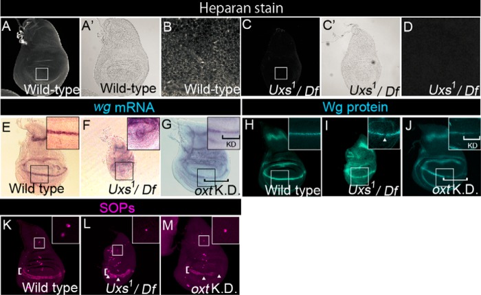
Notch-signaling activation is normal in the Uxs mutant. A–D, wild-type (A, A′, and B) and Uxs1/Df(3L)Exel6112 (C, C′, and D) wing discs stained with an anti-heparan sulfate antibody. A′ and C′ show optic microscope images of A and C, respectively. B and D show magnified views of the white squares in A and C, respectively. E-G, expression of wg, detected by in situ hybridization (magenta) in wild-type (E), Uxs1/Df(3L)Exel6112 (F), and oxt knockdown (G) wing discs. Insets show magnified views of the black squares. Black bracket in G indicates the oxt knockdown region. H–J, distribution of Wg protein, detected by anti-Wg antibody staining (blue) in wild-type (H), Uxs1/Df(3L)Exel6112 (I), and oxt knockdown (J) wing discs; insets show magnified views of the white squares. White arrowhead indicates a gap between Wg protein-expressing cells along the boundary of the dorsal and ventral compartments. White bracket in J indicates the oxt knockdown region. K–M, SOPs detected by anti-Sens antibody staining (magenta) in wild-type (K), Uxs1/Df(3L)Exel6112 (L), and oxt knockdown (M) wing discs. White arrowheads in L and M indicate gaps between Sens-expressing cells along the boundary (white brackets) between the dorsal and ventral compartments. Insets show magnified views of the areas outlined in white, which contain SA neuron precursors. All wing discs were isolated from third-instar larvae cultured at 25 °C except in the RNAi analysis. hh-Gal4 (G and J) or ap-Gal4 (M) was used for the oxt knockdown. All of the oxt knockdown wing discs were isolated from third-instar larvae cultured at 30 °C.

