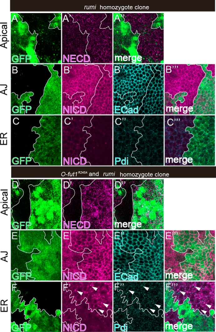
FIGURE 6.

Monosaccharide O-glucose and O-fucose play redundant roles in exporting Notch from the ER. A–F‴, wing discs with somatic clones homozygous for rumi44; A–C‴, or double-homozygous for O-fut1R245A and rumi44; D–F‴ were stained with antibodies against NECD (A′, A″, D′, and D″); NICD (B′, B‴, C′, C‴, E′, E‴, F′, and F‴); DE-cad (B″ and E″); and PDI (C″, C‴, F″, and F‴). Optical images show planes corresponding to the apical membrane (A–A″ and D–D″), AJs (B–B‴ and E–E[tprime]), and the medial region including the ER (C–C‴ and F–F‴). Note that NECD and NICD staining was absent from the apical membrane (D′) and AJs (E′), respectively, whereas NICD staining was increased and co-localized with PDI (white arrowheads in F′–F‴) in the O-fut1R245A and rumi44 double-homozygous cells (lacking GFP). Mosaic clones of mutant cells are indicated by the absence of GFP. Clone boundaries are indicated by white lines. Wing discs were isolated from larvae raised at 25 °C. A″, B‴, C‴, D″, E‴, and F‴ show merged images from A–A′, B–B′, C–C″, D–D′, E–E′, and F–F″, respectively.
