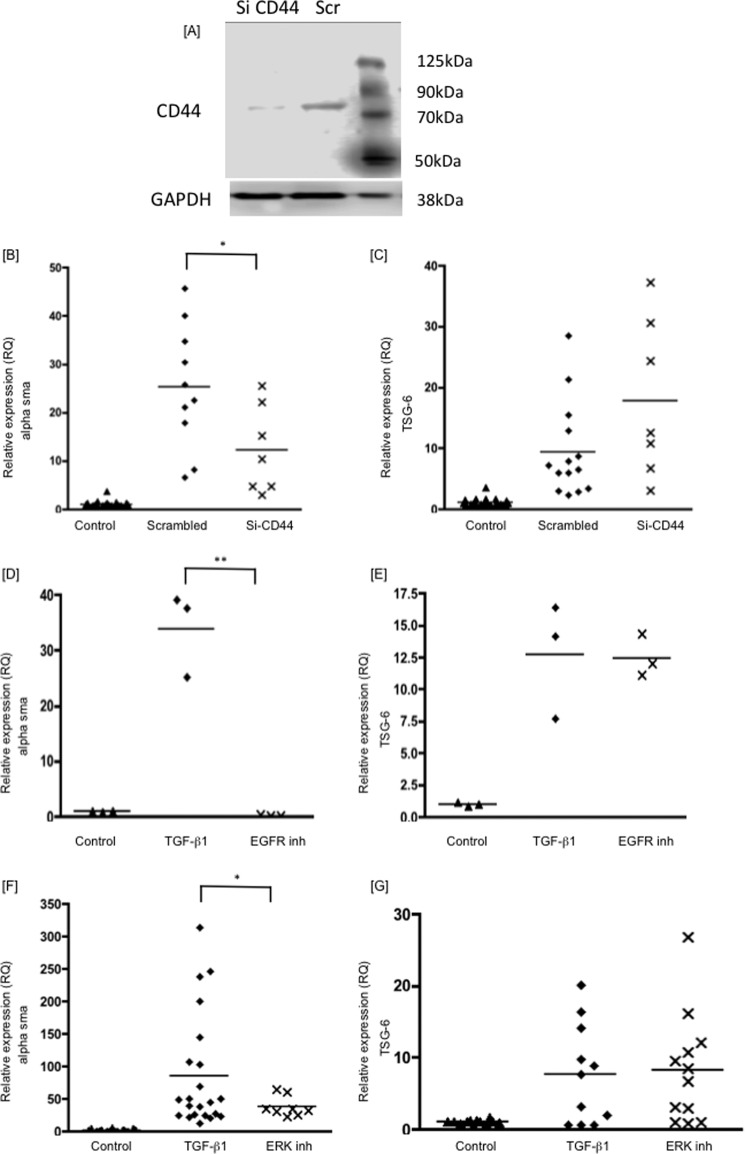
FIGURE 3.
Human lung fibroblasts were growth-arrested and were then stimulated with TGFβ1 (10 ng/ml). The RNA was collected after 72 h of treatment, and the RQ values of α-SMA (B, D, and F), and TSG-6 (C, E, and G) were determined by RT-qPCR. The panels show cells treated with Si CD44, lysed, and protein-extracted for CD44 Western blotting (A); Si CD44 (B and C); EFGR inhibitor AG1478 (10 μm) (D and E); and ERK inhibitor PD98059 (10 μm) (F and G). Inhibitors were added 4 h before TGFβ1 stimulation. Data shown are the mean and scatter of values in three independent experiments, each with between two and three replicates, except one experiment with three replicates in D and E. *, p < 0.05; **, p < 0.005 compared with TGFβ1 treatment alone.

