FIGURE 3.
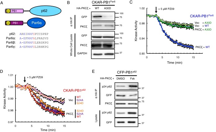
Negative charge at Ser-24/Ala-30 in the PB1 domains of p62 and Par6α impairs binding of PKCζ and activity on CKAR-PB1Par6. A, diagram showing consensus within the PB1 domain for the Ser-24 site of p62 versus the Ala-30 site of Par6α and corresponding Ser sites in Par6β and Par6γ. B, HA-PKCζ was co-expressed with either wild-type (WT) or A30D mutation of CKAR-PB1Par6 in COS-7, immunoprecipitated from soluble lysates using anti-HA antibody, and blotted for co-IP of CKAR tag using anti-GFP antibody, and whole cell lysate was loaded at 10% input. C and D, basal kinase activity of mCherry-PKCζ versus mCherry-Vec on either WT or A30D CKAR-PB1Par6 (C) or WT, S24A, or S24D CKAR-PB1p62 (D) after treatment with 5 μm PZ09 in live COS-7 cells. The trace for each cell imaged was normalized to its t = 0-min baseline value and plotted as means ± S.E. Normalized FRET ratios were combined from four independent experiments (all traces in C and CKAR-PB1p62 S24A Vec trace in D), five independent experiments (CKAR-PB1p62 WT traces and CKAR-PB1p62 S24D Vec trace), or six independent experiments (CKAR-PB1p62 S24A and CKAR-PB1p62 S24D PKCζ traces) with each experiment analyzing 6–13 selected cells. E, HA-PKCζ was co-expressed with WT CFP-PB1p62, treated for 24 h prior to lysis with either DMSO or 50 μm forskolin (Fsk) in COS-7, immunoprecipitated from soluble lysates using anti-HA antibody, and blotted for co-IP of CKAR tag using anti-GFP antibody along with a phospho-specific antibody for Ser(P)-24 p62; whole cell lysate was loaded at 10% input.

