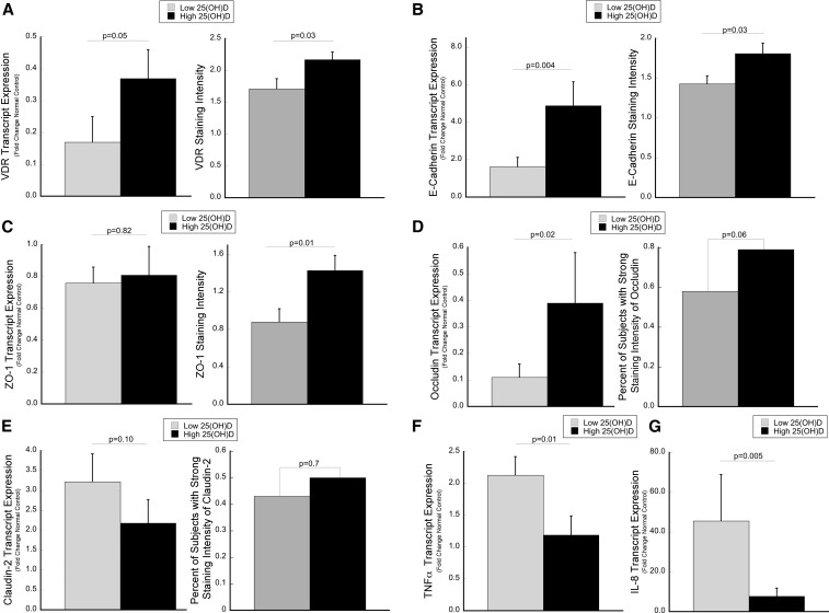
FIGURE 1.
Tissue expression levels of VDR and epithelial junction proteins are increased in association with serum 25(OH)D concentrations, whereas proinflammatory cytokine levels are decreased. Transcript expression by qPCR and protein expression by immunohistochemistry were compared between subjects with low (<20 ng/mL) and high (>25 ng/mL) 25(OH)D concentrations for (A) VDR, (B) E-cadherin, (C) ZO-1, (D) occludin, and (E) claudin-2. (F and G) Transcript abundance of proinflammatory cytokines is shown. The median 25(OH)D concentration within each group was 13.2 (IQR: 10.3–15.8) and 31.9 ng/mL (IQR: 28.7–35.8), respectively, for the transcript analysis and 12.4 (IQR: 10.2–14.6) and 32.8 ng/mL (IQR: 28.3–35.7), respectively, for the protein analysis. For qPCR, a 2-group comparison was made between low and high 25(OH)D concentrations with the use of Student’s t test after each sample was normalized to β-actin with the use of the 2−ΔΔCT method (30). Gene expression was analyzed in 60 subjects, 45 of whom had active inflammation. Protein expression of VDR, E-cadherin, and ZO-1 are presented as the mean staining intensity score, with P values calculated by the Wilcoxon rank-sum test (error bars represent SEMs). Occludin and claudin-2 were scored as strong compared with weak, and P values were calculated with the use of the chi-square test. Protein expression was analyzed in 74 subjects, 54 of whom had active inflammation. qPCR, quantitative polymerase chain reaction; VDR, vitamin D receptor; ZO-1, zonula occluden 1; 25(OH)D, 25-hydroxyvitamin D.

