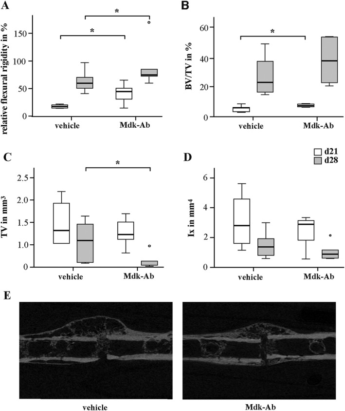
Figure 1.

Mdk‐Ab treatment accelerated fracture healing in mice. Biomechanical and μCT analysis of the fractured femurs on days 21 (d21) and 28 (d28). (A) Relative flexural rigidity of the fractured femur in comparison with intact femur. (B) BV/TV of the fracture callus. (C) TV of the fracture callus at the osteotomy gap. (D) Moment of inertia (Ix) of the fracture callus in bending axis x. (E) Representative μCT images from the fracture calli on day 21. *Significantly different from vehicle (P < 0.05) by Mann–Whitney U‐test. (n = 6–8 per group).
