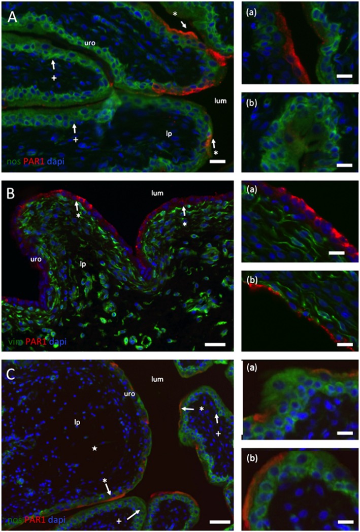
Figure 2.

Location of IR to PAR1 in control (A), CYP‐treated (B) and CYP + F16357‐treated bladders. (A) Sections of the lateral wall of the bladder from a control rat illustrating the regionalized localization of IR to PAR1 (red) to primarily the umbrella cells of the urothelium (*). The lumen (lum), urothelium (uro) and lamina propria are identified. Regions of the urothelium devoid of PAR1‐IR are also apparent (+). The section is counterstained with an antibody to NOS (green) and the nuclear marker DAPI (blue). Calibration bars (A), (B) and (C) 30 mm. (B) Sections of lateral wall from a rat injected with CYP. Note (a) the disruption and loss of the urothelium down to a single cell layer and (b) the appearance of PAR1‐IR in all epithelial cells. No PAR1‐IR was found in the lamina propria interstitial cells. These sections are also stained with an antibody to vimentin (Vim: green) to identify interstitial cells. Calibration bars (B) 40 and 15 mm in (a) and (b). (C) Images from bladders from CYP‐treated animals that also had intravesical F16357 (30 µM). The section is counterstained with an antibody to NOS (green) and the nuclear marker DAPI (blue). Note the absence of urothelial damage and the regional distribution of PAR1‐IR to the umbrella cells. Also, a region of the bladder wall where the lamina propria has expanded (★). Calibration bars (C) 100 mm, (a) 20 and (b) 15 mm.
