
Fig. 4.
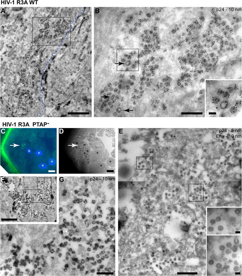
Electron microscopy (EM) immunolabelling of cryosections from monocyte-derived macrophages (MDMs) infected with HIV-1 R3A WT or the PTAP− mutant. Seven-day-old MDMs were infected with HIV-1 R3A WT or the rescued release-defective HIV-1 R3A PTAP−. After a further 7 days, cells were fixed and processed for cryosection immunolabelling. Ultrathin cryosections were immunolabelled with antibodies against p24/55, a fluorescent rabbit-anti-mouse bridging antibody and protein A-gold, or double-labelled for Env as indicated. a, b MDMs infected with HIV-1 R3A WT. Many cells contained intracellular plasma membrane-connected compartments (IPMCs) packed with electron-dense mature virus profiles. The approximate location of the cell surface is indicated by the dashed blue line. Arrows indicate occasional immature viruses (detail shown in the inset). c–g MDMs infected with the HIV-1 R3A PTAP− mutant. c On-grid immunofluorescence staining with the fluorescent bridging antibody (green) shows the location of a cell with IPMC near the corner of grid bars (white arrow). d EM image of the same cell. Note the characteristic pattern of the four nuclei (marked with *) for orientation, indicating the location of the IPMC (white arrow). e A higher power montage image of the boxed area in (d) showing the intracellular membranes with associated immature virus buds (see insets). f A similar view of an IPMC from another experiment. The marked area is shown enlarged in (g). Scale bars: a, 2 μm; b, 500 nm; c, d, 10 μm; e, 1 μm; f, 2 μm; g, 500 nm; and all insets, 100 nm