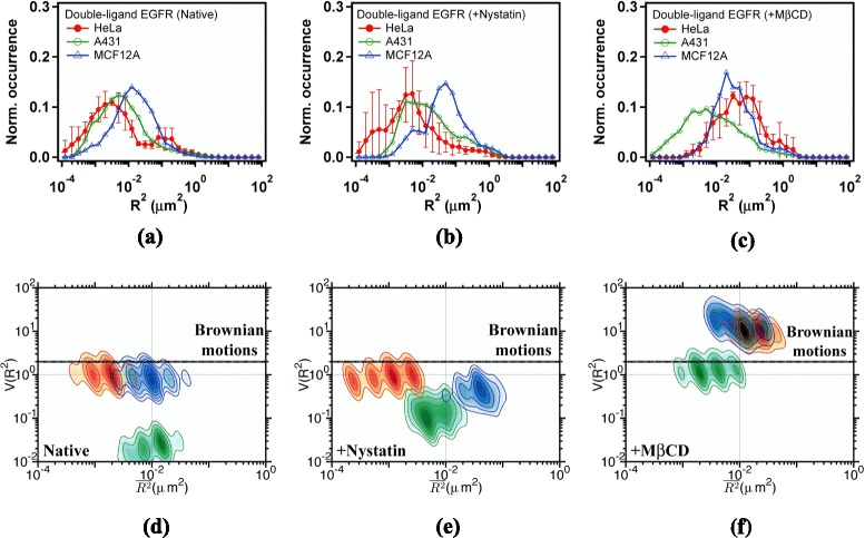
Fig. 6.

a, b, c Histograms of MSDs and (d, e, f) - plots of correlated Qdot585-EGF-EGFRs diffusing in the plasma membrane of (a, d) native cells, (b, e) nystatin-pretreated cells, and (c, f) M βCD-pretreated cells. Trajectory segments with a degree of correlation exceeding 0.8 were analyzed. Trajectories were sampled with a frame period of τ=25 ms. Data are shown in red for HeLa cells, green for A431 cells, and blue for MCF12A cells
