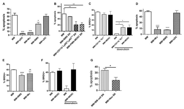
Figure 6. Neutrophils mediate chemoprotective effect on MM through production of soluble factors.
(A) Indicated mouse BM cells were cultured with mouse DP42 cells overnight followed by 24h doxorubicin treatment and detection of apoptosis. (B) DP42 cells were cultured in the presence of supernatants (SN) from BMS, CD11b+Ly6G+Ly6Clow neutrophils or PMN-MDSCs. Cells were then treated with doxorubicin followed by detection of apoptosis. Each experiment was performed independently at least 3 times and the combined results are shown. (C) Proliferation of mouse MM DP42 cells cultured in the presence or absence of CD11b+Gr1+ cells or MDSC for 48h with or without addition of doxorubicin for the last 24h of culture (n=3). (D) Doxorubicin-induced apoptosis of human MM U266 cells cultured in the presence or absence of indicated BM cell populations. (E) Proliferation of U266 cells cultured for 48h with or without indicated cell populations. (F) U266 were cultured with or without MDSCs isolated from BM of 4 different patients with MM and treated with doxorubicin for the last 24h of culture where indicated. Proliferation of MM cells was determined by BrdU incorporation after 48h of co-culture. (G) U266 cells cultured with BM Neu SN or with SN from BM cells depleted of myeloid and MM cells were treated with doxorubicin followed by detection of apoptosis. Combined results from 6 independent experiments performed with SN collected from BM cells obtained from 6 different MM patients are shown. * - p<0.05; ** - p<0.01; *** - p<0.001; **** - p<0.0001.

