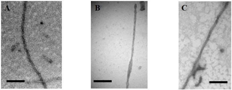
Figure 4.
Electron microscopy of (A) heparin-induced synthetic fibers of Tau441 show the morphology of paired helical filaments, whereas (B) those of a TauF4 fragment devoid of cysteine residues show a morphology of two flat ribbons twisted around one another [126]. This morphology is found in the ex vivo 3R fibers characterizing Pick’s disease [127]; (C) TauP301L after phosphorylation by Erk2 forms fibers without heparin. Scale bar = 100 nm.

