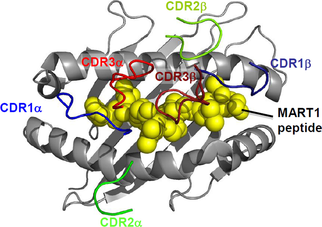
Figure 2. Conserved TCR docking onto pepMHC.
CDR loops of the DMF5 TCR (PDB ID=3QDG) are shown binding to MART1/HLA-A2 as an example of the highly conserved docking geometry observed between TCRs and MHCs. HLA-A2 is shown in gray; MART1 peptide is shown in yellow. CDR3 loops (CDR3α in red, CDR3β in brick red) dock predominately over peptide, while CDR2 loops (CDR2α in green, CDR3β in lime green) predominately contact MHC. CDR1 loops (CDR1α in blue, CDR1β in navy) can contact both the MHC and the peptide; the CDR1α loop in particular frequently makes specific contacts with the N-terminus of the peptide.

