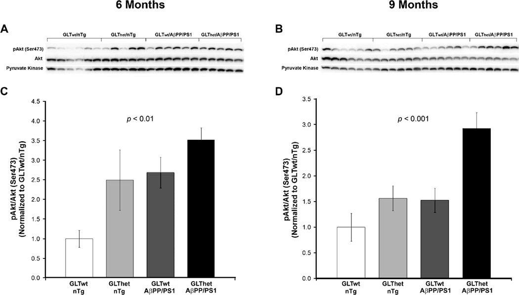
Fig. 1.
Akt phosphorylation is increased by partial loss of GLT-1 and expression of the AβPP/PS1 transgene and is more pronounced at 6 months of age. A, B) Western blots from 6 and 9 month animals, respectively. C, D) Indicating sustained activation of Akt, densitometric quantification of blots in (A) and (B) revealed a statistically significant increase in the ratio of phosphorylated Akt (Ser473) to total Akt in GLThet and AβPP/PS1 transgene mice compared to GLT-1wt/nTg mice at both 6 (C) and 9 (D) months of age (p ≤ 0.01 and p ≤ 0.001, respectively). Each lane represents one animal. Data are presented as normalized means ± SEM (n = 5–6 mice per group).

