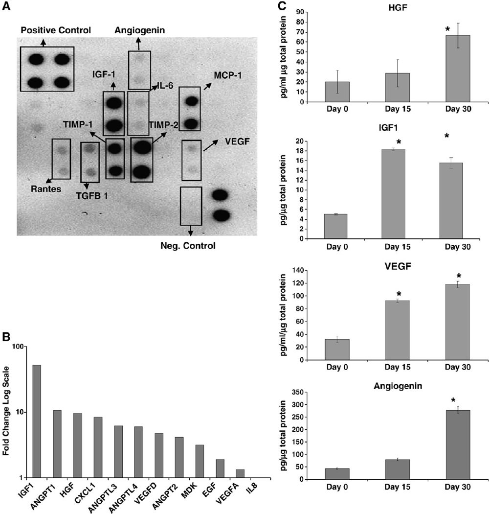
Figure 2.
SD-MSCs express angiogenic factors: (A) Immunoblot of culture media conditioned by SD-MSCs on Day 30 of serum deprivation. (B) Relative expression of mRNA of secreted angiogenic factors in SD-MSCs from Day 30 of serum deprivation compared to donor-matched MSCs, as determined by real-time PCR. Fold change in transcript levels is represented on the Y axis in a log scale. (C) ELISAs of growth factors in conditioned media from MSCs (Day 0) and SD-MSCs (Day 15 and Day 30). Values below the minimum detectable level of the assay are marked with a #. The average of 4 culture replicates with SD is shown. * indicates a significant difference from Day 0 values (P < 0.01).

